Collapsed lung with Empyema
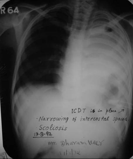
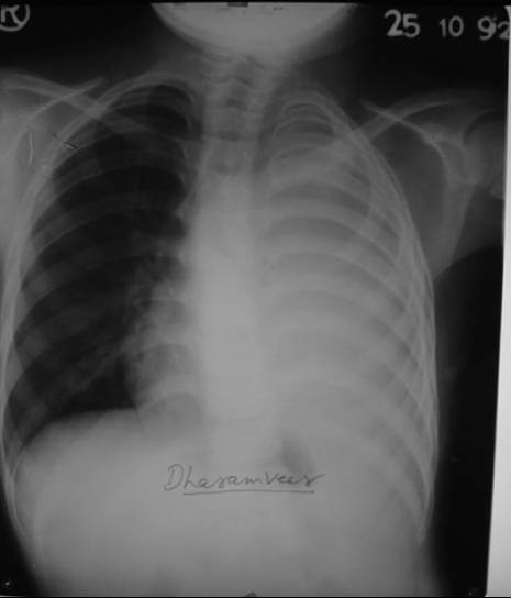
Presenting complaint and history On 6 October 1992, a 10 year-old male presented after two convulsions the previous day and a discharging sinus in left chest. He was admitted on 10 august 1992 complaining of fever with chill, left-sided chest pain on coughing and on mild exertion, cough dyspnoea and loss of appetite for 15 days. After clinical examination and full investigation the case was diagnosed as collapsed lung with tubercular empyema Anti tubercular treatment (ATT) was started with broad-spectrum antibiotics and regular drainage of the pleural cavity (fig1) but the condition deteriorated (fig 2) Dated: 11 Aug. 1992 See the intercostal drainage tube in place. Dated: 23 Aug. 1992. Tense pyothorax on removing the intercostal tube. Both x-rays (fig.1 and fig.2) are taken while the patient was under care of modern system of medicine. After few days of ATT, jaundice appeared and this was followed by the convulsions. He was referred to the cardiothoracic surgeon for decortication, but the surgeon decided not to operate because he had not responded to the ATT. At this stage his parents decided to discontinue the ATT and asked for homeopathy. After evaluating the history it was obvious the convulsions arose as a complication of ATT and untreated empyema. I started the homeopathic treatment with few doses of phos 30. He had no more convulsions since first dose of homeopathy.
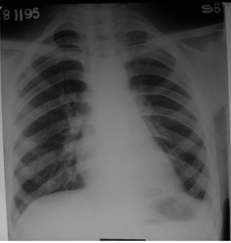
This case improved gradually and gets cured within six months. Till date, he has no problem.
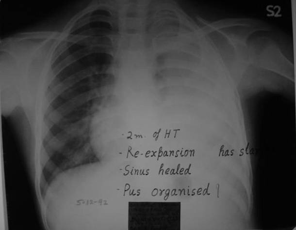
Dated. 5 Dec.1992.
Re-expansion had started after one month of homeopathic treatment.
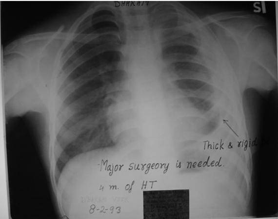
Dated: 8 Feb.1993.
Re-expansion of collapsed lung was in progress. Sinus had healed.
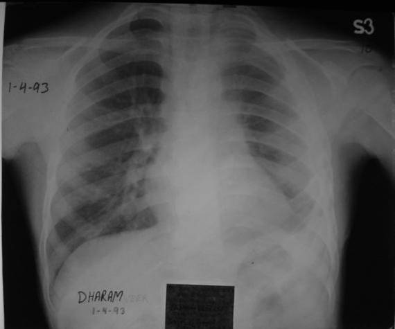
Dated: 1 April 1993.
See the gradual non-stop re-expansion of lung. Pleura had become thick and fibrosed.
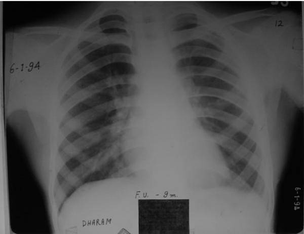
Dated: 6 Jan. 1994.
Again there is a process of resolution of fibrosed pleura is visible.
Dated. 5 Dec.1992.
Re-expansion had started after one month of homeopathic treatment.
Dated: 8 Feb.1993.
Re-expansion of collapsed lung was in progress. Sinus had healed.
Dated: 1 April 1993.
See the gradual non-stop re-expansion of lung. Pleura had become thick and fibrosed.
Dated: 6 Jan. 1994.
Again there is a process of resolution of fibrosed pleura is visible.
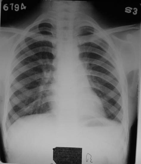
Dated: 6 July 1994. Follow
-up normal x-ray
Discussion:
Pus in long standing Empyema becomes organized and fibrosed leads to immobilization of the affected side of lung. As per Medical Literature Fibrosis can not be reverted but it is not so in this case. Fibrosed and thickened pleura ultimately gets normal in its shape.
Pus in long standing Empyema becomes organized and fibrosed leads to immobilization of the affected side of lung. As per Medical Literature Fibrosis can not be reverted but it is not so in this case. Fibrosed and thickened pleura ultimately gets normal in its shape.
